This state-of-the-art MRI machine produces better, faster, and less expensive images of the human body, thus improving the capability of diagnostic imaging to detect diseases.

By Justin Chapman
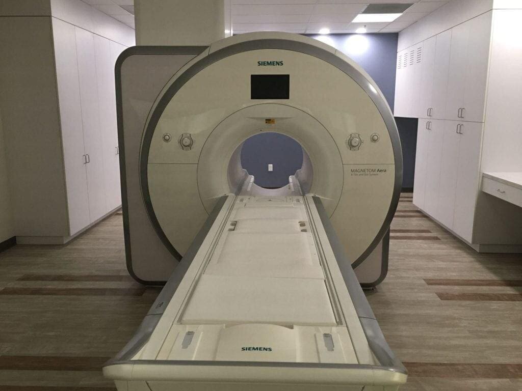
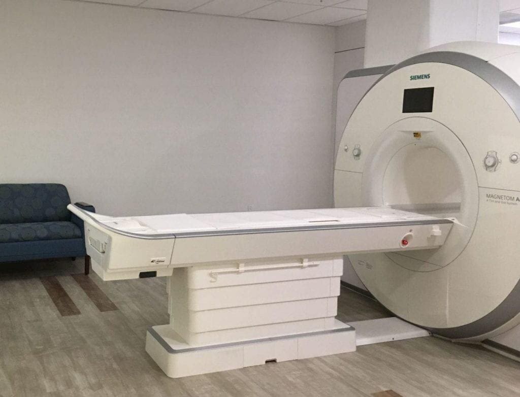
The state-of-the-art magnetic resonance imaging (MRI) machine that was recently installed in the USC Michelson Center for Convergent Bioscience (MCCB) allows researchers and students to produce better, faster, and less expensive images of the human body, thus improving the capability of diagnostic imaging to detect diseases.
As Ben Paul of the USC Viterbi School of Engineering explained, “Traditional MRI machines struggle to produce reliable images of certain body parts, such as the heart, lungs, and vocal tract, and of body parts that are near metallic implants. They are also exceedingly large and expensive.”
The low-field, high-resolution, high-performance 0.55 Tesla whole-body MRI scanner is one of only three of its kind in existence and was installed in December by an initiative led by Dr. Krishna Nayak, Viterbi professor of Electrical and Computer Engineering. The machine was installed in Viterbi’s newly established Dynamic Imaging Science Center (DISC), which officially launched in May with support from the National Science Foundation.
Built by Siemens, the MRI scanner has a “wide range of physiological monitoring systems that can be utilized simultaneously with imaging,” according to DISC’s website. “Current projects include the imaging of the upper airway vocal tract during speech production, swallowing, and sleep; imaging movement of joints such as the wrist; and cardio-pulmonary imaging.”
“We use diagnostic imaging extensively in medicine. It’s one of the reasons we’re able to offer personalized treatments, therapies, and assessments. We’re trying to develop the best possible diagnostic tools for diseases.”
—Dr. Krishna Nayak
DISC’s mission is to “better understand the science of human movement in health and disease, through the development and use of non-invasive imaging.” The Center is supported by the National Science Foundation, the USC Viterbi School of Engineering, and Siemens Healthineers.
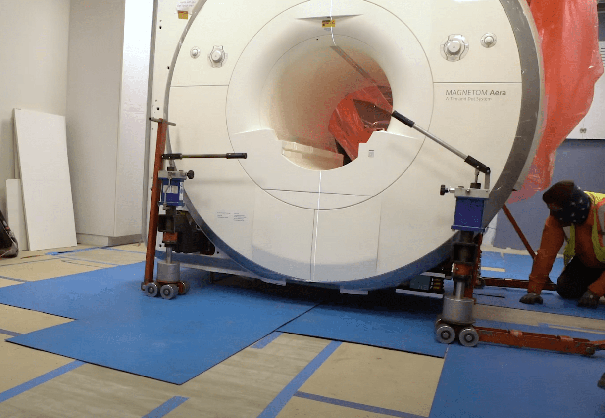
The MRI machine, which weighs 4.8 tons, was lowered into the building on a crane and rolled down the hallway into place. Watch a video of the installation here. MCCB is USC’s flagship biomedical research building.
With traditional MRI machines, patients must lay still for anywhere from 30 minutes to an hour for the course of the MRI exam. This MRI machine is much more versatile.
The new machine “allows us to get superior performance in areas of the body that are limited by artifacts, and that’s why we are focusing on imaging around post-surgical patients, orthopedic metallic implants, parts of the body that move, or the unborn fetus,” Nayak said. “These are all diagnostic imaging challenges, where the conventional field strengths are not optimal.”
The machine’s field strength, 0.55 Tesla, is three times lower than the most common MRI system and six times lower than the system that’s predominantly used at USC. That results in a loss of image quality but allows researchers to view certain areas of the body such as the lungs and heart that are obscured by various artifacts.
“Low” field strength is relative, he added, as the magnetic field strength is still about 10,000 times stronger than the earth’s magnetic field.
“If you have a healthy brain with no metallic implants or wires, then the lower field strength is going to be a huge penalty in quality,” Nayak said. “I don’t expect this scanner to ever be used for high-end neuroscience research, but if you have a patient that has had a brain tumor and had to have a craniotomy and they have a metal plate on their skull, you’re not limited by the signal, you’re limited by the artifacts from that metal plate.”
Nayak explained that the new technology will have a big impact on a wide range of disciplines.
“We use diagnostic imaging extensively in medicine,” Nayak said. “It’s one of the reasons we’re able to offer personalized treatments, therapies, and assessments. We’re trying to develop the best possible diagnostic tools for diseases.”
Wide-Ranging Applications
Researchers from a number of different schools at USC, including Viterbi, Dornsife, Keck School of Medicine, Children’s Hospital of Los Angeles, and more will be able to utilize this new technology. Researchers from different disciplines say the new MRI machine will help them in their individual fields in different ways.
“Cardiologists tell us that they need to have a better assessment of patients with cardiac arrhythmia or people that have implanted pacemakers or defibrillators,” Nayak said. “Being able to scan them without having to reprogram their device would result in healthcare cost savings.”
For instance, fetal imaging specialists could use MRI more often to detect brain and cardiovascular abnormalities.
When it comes to the lungs, the new MRI machine can potentially detect lung cancer or COPD early. It can also detect failures of orthopedic metallic implants and provide images of the soft tissue around those implants.
“It’s also bucking a current trend in imaging, which is to go to higher field strengths which are very expensive,” Nayak said. “So if we’re able to make the system work, another potential community benefit is that this could be much more cost effective than other imaging instruments.”
“We use diagnostic imaging extensively in medicine. It’s one of the reasons we’re able to offer personalized treatments, therapies, and assessments. We’re trying to develop the best possible diagnostic tools for diseases.”
—Dr. Krishna Nayak
Nayak added that Siemens has recently announced that they’re going to make one of their next generation MRI machines at this field strength, 0.55 Tesla.
The machine more reliably handles patients with implants in a much quieter environment and has the potential to bring MRI technology to communities and facilities that cannot afford traditional MRI machines, according to Paul.
“This is excellent for rural areas, where it’s often complicated to site an MRI system,” Nayak added. “So there could be a huge improvement in access to this capability, which is currently a big problem. High-end diagnostics are not really available everywhere in America or around the world.”
In addition to lead principal investigator Nayak, the project has four co-PIs: USC Viterbi faculty Dr. Shrikanth Narayanan and Dr. Justin Haldar and USC Dornsife faculty Dr. Dani Byrd and Dr. Khalil Iskarous.
Nayak’s research group at MCCB recently published a paper in Scientific Data about their research using the machine for real-time magnetic resonance imaging (RT-MRI) of human speech production, which is enabling significant advances in speech science, linguistics, bio-inspired speech technology development, and clinical applications.
“The complexity and sophistication of human speech production poses a multitude of open research questions with implications for linguistics and speech science, as well as clinical and technological applications, creating a demand for improved methods for observing and measuring the vocal instrument in action,” they wrote. “RT-MRI with concurrent audio recording has emerged as an imaging modality that can provide new insights into speech production with all its inherent systematicities and variation between languages, contexts, and individuals.”
“Low-field MRI might have an even larger economic impact on society in the future. The weaker magnet used in low-field MRI means these machines could cost much less and become much smaller and easier to maintain. The machine’s home in Michelson Hall is appropriate, as it represents a true convergence of engineering and medicine.”
—Ben Paul
Their paper presented a unique dataset including raw MRI data and videos of the entire vocal tract in action, with synchronized audio, imaged along the sagittal plane—an anatomical plane which divides the body into right and left parts—from 75 participants while they performed a variety of speech tasks. Their study can result in improved models of speech production in linguistics and speech science research.
“It is our hope and anticipation that the public and free provision of this rich dataset can further foster and stimulate research and innovation in the science of human speech production and its imaging,” they wrote.
Nayak’s group includes three Ph.D.-level scientists and 10 Ph.D. students. Five faculty members actively use the MRI machine to scan people on a regular basis. An additional 25 faculty members are collaborators on projects and refer patients or assist with data analysis. The machine is also being used to train a number of Ph.D. students, and will soon be included in undergraduate and graduate courses.
“We’re in this rapid growth phase, but it’s primarily the technology-heavy users using the MRI machine right now because we have to get everything working for the first time on this field strength,” Nayak said. “We’re keeping it tight in the beginning because of COVID. When we are able to have more of a variety of people coming in, by December, we’ll be scanning patients and it will open up quite substantially.”
In a Viterbi Magazine article, Paul wrote that “low-field MRI might have an even larger economic impact on society in the future. The weaker magnet used in low-field MRI means these machines could cost much less and become much smaller and easier to maintain. The machine’s home in Michelson Hall is appropriate, as it represents a true convergence of engineering and medicine.”
In 2014, a $50 million gift from Dr. Gary Michelson and his wife, Alya, established the USC Michelson Center for Convergent Bioscience, which brings together exceptional scientists, researchers, and engineers from around the world to work collaboratively on solutions to today’s most pressing health issues. This 190,000-square-foot interdisciplinary research facility is a model for other research universities, bringing the world closer to the medical breakthroughs that are redefining the 21st century.